In 1834, the year in which Charles Darwin’s HMS Beagle reached the west coast of South America, slavery was officially abolished throughout the British Empire and Mount Vesuvius erupted, the London Times ran a short article on the stethoscope, then a relatively new medical device to hit the market. It contained this quote: "That it will ever come into general use, notwithstanding its value, is extremely doubtful; because its beneficial application requires much time and gives a good bit of trouble both to the patient and the practitioner; because its hue and character are foreign and opposed to all our habits and associations."
History has, of course, laid waste to this antiquated argument, although medical developments have – in many cases, understandably – continued to court such reactionary responses.
Go deeper with GlobalData

In 1988, 154 years after the publication of the London Times piece, Dr Roy Filly, then chief resident at the faculty of radiology at the Stanford University School of Medicine issued a similar rebuttal concerning the use of diagnostic sonography. In his report entitled ‘Ultrasound: the stethoscope of the future’ featured in Radiology, he argued: "As we look to the proliferation of US [ultrasound] instruments into the hands of untrained physicians, we can only come to the unfortunate realisation that diagnostic sonography truly is the next stethoscope: used by many, understood by few."
Filly’s words carry greater prescience. Once the sole preserve of radiology departments, the use of ultrasound for procedure guidance, diagnosis and screening has proliferated dramatically in recent years and is presently used in a number of medical specialities.
Common uses of sonography
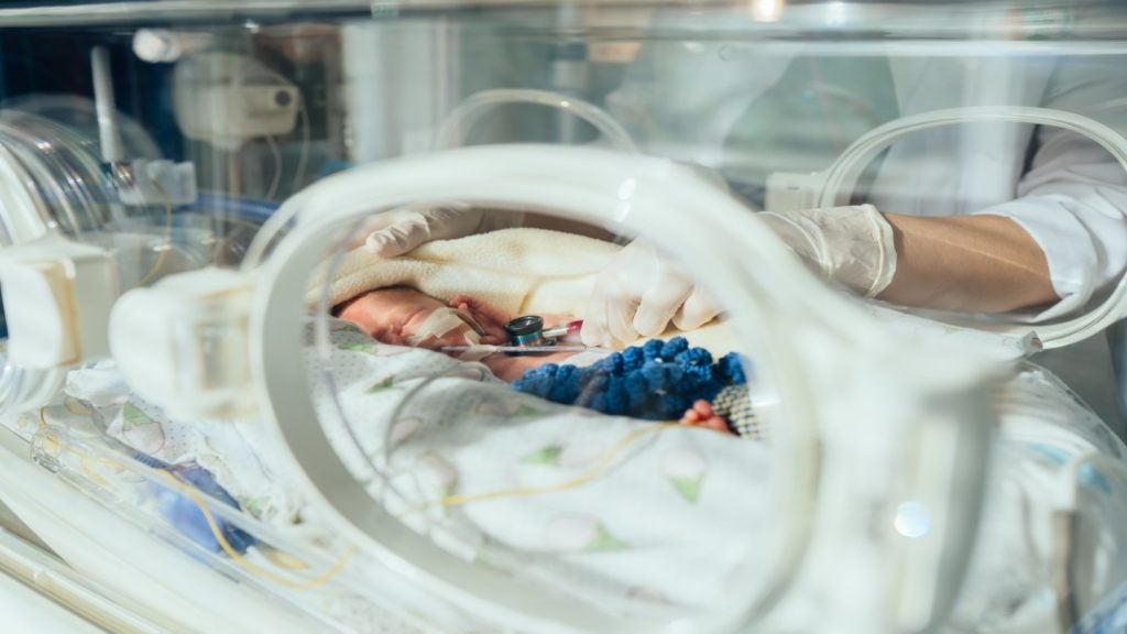
Perhaps best known for its use in prenatal care, sonography is commonly used by clinicians – ranging from trauma surgeons to obstetricians and cardiologists – in screening for stones, ectopic pregnancy, vascular access and bladder volume measurement.
See Also:
According to Deborah Levine, chair of the American College of Radiology Ultrasound Commission and professor of Radiology, Harvard Medical School, the trend has been in full swing for some time now.
How well do you really know your competitors?
Access the most comprehensive Company Profiles on the market, powered by GlobalData. Save hours of research. Gain competitive edge.

Thank you!
Your download email will arrive shortly
Not ready to buy yet? Download a free sample
We are confident about the unique quality of our Company Profiles. However, we want you to make the most beneficial decision for your business, so we offer a free sample that you can download by submitting the below form
By GlobalData"Specialities other than radiology have been interested in ultrasound for a while," she says. "And as the machines have become smaller and more portable, particularly over the last decade, it has become easier for these subspecialities to be able to get less expensive machines, without all the bells and whistles on, in order to answer very specific questions."
The advantages of point-of-care appear to be writ large: machines are easy to use, small, portable and safe, and procedures do not use ionising radiation. Costs are also relatively low compared with those found with other techniques, such as magnetic resonance imaging and computed tomography.
Furthermore, because procedures are conducted at the bedside, patients are availed of greater comfort; images can be obtained almost immediately, and clinicians also have access to real-time images, as opposed to them being sent away with sonographers to be analysed later.
Point-of-care systems: proliferation and limitations
However, Levine believes there has been something of a trade-off between the modality expertise of more complete examinations conducted by radiology departments and the speed of diagnosis found with new point-of-care systems.
"Point-of-care ultrasound might offer a faster diagnosis, but I think the high-end machines used by radiology departments are still the final word when it comes to better resolution and sensitivity," she says. "So, there’s always that compromise, but it really depends on what you need the machine for."
In February last year, the New England Journal of Medicine (NEJM) published a report, ‘Point-of-care ultrasonography’, in which its authors reviewed the current proliferation of equipment. And while attributes such as efficiency and accuracy of portable devices were extolled – there is currently an accuracy rate of 90-98% found across emergency diagnoses – question marks still remain over user competence.
So what levels of training are on offer to care providers and clinicians?
"That’s a question that is actually very problematic to me as a radiologist that specialises in ultrasound," admits Levine. "While, here at the ACR, technologists are required to go to sonography school, there are currently no exams for point-of-care procedures. So there is no minimum standard – anyone can procure one of these machines and start using them with no training at all."
Echoing Filly’s earlier words, Levine argues that, in effect, point-of-care ultrasound is in danger of marking a significant step backwards, with many clinicians returning to learning on the job, as opposed to setting aside time to receive the appropriate training.
"We have seen a return to the days of on-the-job training, which is something I find slightly worrying," she says. "Even if the machine has all these amazing capabilities, the person holding the probe might not know how to get best images or have a sufficient understanding of how to use the different knobology functions."
Anomalies and false positives
Technology cannot come at the expense of qualified expertise, with ultrasound diagnoses and critical decisions ultimately being contingent on physicians’ knowledge and ability. This is particularly pertinent in the case of anomalies and false positives – results indicating that a given condition is present when it is not.
"Both false positives and false negatives are common," says Levine. "For instance, in the case of ectopic pregnancy [a complication in pregnancy where the embryo implants outside the uterine cavity], if you don’t have enough experience, it is quite easy to diagnose incorrectly.
"Likewise, when inspecting a patient with right upper quadrant pain, it is a widespread mistake to concentrate too much on finding gallstones and neglect possible abnormalities in the liver. So, even with the best of intentions, improper diagnoses can still be made."
With such pitfalls still to be ironed out, it is unlikely that point-of-care is set to replace more sophisticated systems and rigorous studies conducted by radiology departments any time soon. Patients with more complex conditions will continue to be evaluated on high-end consoles as part of complete examinations.
However, as technology advances at an inexorable pace, so point-of-care will continue to afford more options to medical staff and patients. Last year’s NEJM report also cited the new-found potential of point-of-care for detecting the likes of pneumothrax – commonly known as collapsed lung – a condition previously deemed beyond the reach of ultrasonography.
In Levine’s eyes, such advancements only throw the need for training procedures and quality assurance into greater relief. In addition to mandatory training, she would like to see point-of-care images and reports made available to other clinicians, which she argues is "currently not part of the culture".
Need for greater quality assurance
"I would like to see more quality assurance mechanisms in place," she says. "In other words, images are stored so that other clinicians can look at them and check whether diagnoses are correct. This would entail the documentation of images as well as written reports, which is something you have with radiology department where images are either stored in a film library or on a computerised system such as a PACS [picture archiving and communications system]."
And what of the argument that the increase in point-of-care ultrasound is commensurate with indiscriminate and inappropriate use by physicians?
It’s hard to say, but the positives appear to far outweigh the negatives. In recent years, portable ultrasound systems have also had a huge impact beyond hospital confines, coming into play in more austere and remote environments such as war zones and natural disaster areas. Notably, in the aftermath of the 2010 Haiti earthquake, scores of victims were able to benefit from remote sonography used by emergency response teams.
This potential to save lives and protect patients is ultimately the nub of the matter, as Levine explains: "When it comes down to it, the most important thing we need to focus on is the optimal care of the patient," she says.
With additional knowledge, training and improved critical decision-making, any reservations over point-of-care ultrasound may one day be consigned to the past, just as similar misgivings over the stethoscope gave way many moons ago.