Medical education in the US has undergone dramatic changes in the past decade. One reason for this has been to provide efficient and effective medical education in an environment that requires multidisciplinary approaches to diagnosis and treatment for optimal patient care.
For example, the first year anatomy and radiology course at Mayo Clinic College of Medicine, US, has been transformed into an accelerated six-week combined course that includes (laboratory-based) gross anatomy, embryology and an introduction to radiology. The radiology instruction in the course includes use of interactive 3D visualisation of CT scans of each cadaver at the dissection table and one-on-one contact with clinical anatomists and radiologists in the anatomy laboratory. The incorporation of technology and advanced clinical tools into this new course has been key to the students’ assimilation of the radiology and anatomy course materials.
Go deeper with GlobalData

Old school
Traditionally, medical school training consists of an early focus on ‘basic science’ elements including separate classroom-based coursework in anatomy, histology, embryology, organ-system physiology and relevant biochemistry. Anatomy has arguably the longest history as a component of formalised medical education, and is nearly always a required part of medical school education.
Later coursework typically includes application of this knowledge to the study and treatment of disease including pathology, pharmacology, microbiology and immunology. The application of this basic knowledge to actual diagnosis and patient care traditionally occurred in the later portions of medical school training, when clinical clerkships, physical examination and other hands-on skills are taught through formal coursework, apprenticeship/mentorship-like exposure and independent study.
See Also:
Until recently, the gross and developmental anatomy course at the Mayo Clinic College of Medicine had been taught in a relatively traditional curriculum: a five-month course as part of a basic science curriculum during the first year of medical training. This course included 50 hours of classroom lectures and clinical correlations and about 130 hours of hands-on dissection of cadavers by medical students (working in groups of four) with faculty and teaching assistants (third-year medical students) supervising.
How well do you really know your competitors?
Access the most comprehensive Company Profiles on the market, powered by GlobalData. Save hours of research. Gain competitive edge.

Thank you!
Your download email will arrive shortly
Not ready to buy yet? Download a free sample
We are confident about the unique quality of our Company Profiles. However, we want you to make the most beneficial decision for your business, so we offer a free sample that you can download by submitting the below form
By GlobalDataAlthough the clinical anatomy, peer-teaching, elements of teamwork, leadership, and professionalism were incorporated into this curriculum, the anatomy course was independent from the other first-year basic science courses. This is typical of a traditional US medical school anatomy course, where the first exposure to radiology and medical imaging occurs during the semester-long anatomy course taught by an anatomist.
In Mayo’s traditional anatomy course, the appearance of anatomic structures on radiographic studies was used to make educational points and provide clinical relevance; however, the imaging instruction was not taught in collaboration with radiologists and there was no contiguity between radiographic anatomy education and the radiology course. The previous radiology course was taught later in the curriculum, just before students began their clinical clerkships. This radiology course was focused, intense, and included discussion of the different types of imaging modalities used in modern radiology:
- X-ray and X-ray fluoroscopy (and current digital radiography and computed radiography equivalents)
- computed tomography (CT)
- magnetic resonance imaging (MRI)
- nuclear medicine positron emission tomography (PET)
- ultrasound.
The appearance of normal anatomy and pathology on these different types of studies was emphasised, but the radiology curriculum was independent from both the formal anatomy and pathology courses. This brief course was the only direct radiologic instruction received by most of the students during medical school.
Teaching methods
The methods used in teaching, medical skills acquisition and presentation of basic science materials should be combined with relevant clinical examples. Similarly, hands-on interaction with clinical tools and one-on-one interaction with practising clinicians during what would be considered a traditional basic science course enhances the educational experience. Specifically, traditional basic science courses are improved by being streamlined and combined with case-based clinical content and hands-on practical skills development.
Such multidisciplinary approaches to medical education may overcome the disconnect between various components of the learning process and better clarify learning objectives, facilitate teaching and augment retention of knowledge in practice. It has been previously demonstrated that the use of electronic systems for distribution of educational content, inclusion of new methods of content representation, or visualisation by computers can supplement traditional methods of education and enhance outcomes.
Specifically, for anatomy and radiology education, a multidisciplinary approach using innovative technology to convey knowledge and anatomic relationships with clinical imaging tools should improve the ability of students to recall and expand their base of anatomical knowledge throughout their medical school training and beyond the span of the anatomy course to subsequent course material.
3D visualisation tools
Medical imaging technology has recently shown tremendous advances with the application of new and better tools for diagnosis, surgical planning, treatment and monitoring of disease in response to therapy. In clinical practice, use of 3D visualisation allows for enhanced diagnosis, accurate treatment planning and rehearsal, and education/training. It has been shown that simulation and use of interactive 3D imaging can be a useful training aid for surgeons.
Interactive 3D volume rendering can also help students overcome many of the conceptual limitations encountered when conveying or teaching 3D relationships via traditional diagrammatic representations or 2D imaging. Even more complicated relationships, such as the relative position and changes in relative position of cardiac and coronary artery anatomy during the cardiac cycle, can be more usefully depicted with interactive 3D and 4D (including the dimension of time) modeling. Use of interactive volumetric renderings may even be necessary for acquiring surgical skills. This also aids in the understanding of the perceptual constraints involved in minimally invasive surgery training.
A thorough understanding of anatomy through hands-on dissection and early coursework remains an essential part of medical education. However, there can be improved dissection efficiency in the human gross anatomy laboratory by the integration of computers, particularly when imaging studies available at the dissection table depict the actual anatomy of the cadavers being dissected.
The increasing sophistication with which modern imaging can depict anatomy, combined with web-based electronic availability of interactive 3D volumes that demonstrate this anatomy, means that radiologists and anatomists can collaborate to improve the efficiency of acquisition and retention of anatomic knowledge for tomorrow’s physicians.
Familiarity with these advanced visualisation tools early in training enables more efficient learning of complex anatomy and the interrelationship of anatomic structures. Early exposure to the discipline of radiology allows for the transfer of clinically relevant knowledge to students from radiologists with subspecialities during basic science coursework.
Clinical radiology examinations
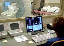
In clinical radiology, practice examinations are typically viewed on dedicated high-performance medical imaging devices. For better visualisation of some structures and the relationships of pathology to anatomic structures, multi-planar (axial, sagittal, coronal or oblique) reformatted displays or 3D renderings of the thin slice data can be performed.
Due to the size and complexity of many digital radiology examinations, the medical devices that can perform these operations quickly and efficiently for medical practice are typically not portable nor can the software that makes the rendering be used on an ordinary PC. In the anatomy laboratory where students perform their dissections, standard PC laptop computers are used that are connected wirelessly to electronic course content and reference materials.
To enable access to the CT data for each cadaver and interactive volumetric 3D renderings of these large and complex datasets, an innovative web-based, thin client display software was used (ViTALConnect® software version 4.0). This software is a web-enabled medical diagnostic tool, which in clinical practice, allows physicians to use PCs or notebook computers to access interactive 2D, 3D and 4D advanced visualisation.
A majority of the medical imaging modalities can be viewed using the software and are not limited to CT, MRI, CR, DR, NM, PET or ultrasound.The software is similarin functionality to dedicated 3D workstations, but as a thin client/server solution it offers clinical users access to advanced visualisation that is critical in today’s distributed hospital environment.
To facilitate knowledge of cutting-edge imaging technology as part of the anatomy/radiology course, each cadaver was scanned from head-to-toe at high resolution with a modern multislice CT scanner. Images of the cadavers as well as normal references images, including plain radiographs, MRI scans, nuclear medicine studies and ultrasounds, were used during the didactic portion of the course to demonstrate radiologic/anatomic correlations.
Interactive access to the reference image data and the CT scan data of each cadaver was available to students at each dissection table through the use of web-based software that allows colour 3D renderings to be distributed from a central rendering server (ViTALConnect).These images were streamed to laptops in the anatomy laboratory, which allows for visualisation of specific structures encountered during dissection much as they would be seen in a living patient with a clinical CT scan.
This database of reference radiology datasets and cadaveric visualisations are particularly useful in appreciating pathology observed in the specific cadaver being dissected and for understanding the complex relationships of anatomic structures in space. Similarly, the web-application’s images can be stored in other electronic formats (jpg, tif and avi), directly saved, printed or inserted into other applications. Students can include selected radiologic images in the required ‘autopsy reports’ that they prepare at the end of the course.
Physicians of tomorrow
Innovative methods of medical education must be used to instill the knowledge and skills necessary for future physicians. Redesigning the curriculum at the Mayo Clinic College of Medicine opened an opportunity to create a new educational framework for anatomy that incorporates radiology instruction into first-year medical student education.
Although some introduction to radiology has been part of traditional anatomy education for decades, the expanded role of imaging in both medical care and relevance to anatomy education provides opportunities for exposure to radiologic imaging early in medical education and use of imaging to support the learning of anatomy and the basic sciences.
Specifically, the one-to-one correlation of anatomy and 3D visualisations of cadaver CT scans allows for better understanding of the anatomy and pathology apparent on clinical radiology studies used for patient care. Cutting-edge technology allows students to access and interact with the CT data of their own cadaver and other reference radiological images at the tableside during dissection as interactive 3D volumetric colour renderings.
Real-time volumetric visualisation tools and radiologic studies during dissection allow for concurrent correlation of the radiographic appearance directly with the anatomy and pathology. Through this visualisation system, students are given the opportunity to correlate their observations during dissection with a radiologic image of their choosing.
The intent of the course structure is to help the next generation of medical practitioners appreciate what can be seen in a patient through radiologic examinations and provide immediate comparison between radiology imaging and what is seen during traditional dissection.
The exposure of medical students to modern clinical imaging techniques allows for early familiarity with these powerful imaging tools that will certainly be commonplace in clinical practice in their future careers. It is hoped that these innovations in medical education enable efficient education, and that the next generation of physicians has both the knowledge and clinically relevant skills to practise in an environment that increasingly relies upon sophisticated medical imaging for diagnosis, surgical planning and disease management.
References available from the authors on request.