
Breast cancer remains a major health problem for women despite significant advances in the treatment of cancer. In 2008, approximately 45,000 women were diagnosed with breast cancer in the UK, and over 12,500 will die as a result; in the US, about 180,000 women were diagnosed with breast cancer, of which 40,480 will die. In the UK, women have a one-in-nine risk of developing breast cancer, and in the US the risk is one in eight.
An important part of improving the chances of successful breast cancer treatment is the early and accurate detection of clinically significant changes in the breast. Until recently, the primary means of detection has been through X-ray mammography and ultrasound imaging.
Go deeper with GlobalData

Discover B2B Marketing That Performs
Combine business intelligence and editorial excellence to reach engaged professionals across 36 leading media platforms.
Detection methods
Dr Carrie Hruska and a team of researchers at the Mayo Clinic in Rochester, MN, US, are working on molecular breast imaging (MBI), a new imaging technique that promises to improve the quality of detection and, hopefully, increase the numbers of patients that are treated successfully.
“The standard of care for women who are asymptomatic, without any breast problems, is routine screening via an annual mammogram,” says Hruska. “In the US, that would be for all women over 40. For many years film mammography was standard, but that is quickly changing to digital, in the US at least.”
Additional detection methods are used in conjunction with mammography – the most common being ultrasound, which is typically used as an additional diagnostic tool to take a closer look at something that has shown up on a mammogram. Ultrasound has a valuable role because it can be used to, for example, examine a lesion to show whether it is benign or malignant. Using ultrasound guidance, it also allows for an easier biopsy.
In addition to mammography and ultrasound, another level of care is magnetic resonance imaging – the breast MRI. But, while MRI is becoming more widely used in the US and Europe, it is usually used in situations where something suspicious appears on a mammogram or ultrasound that cannot be resolved or as a screening technique for women with a very high risk of breast cancer. MRI is more complicated, more costly to the patient and, although it is has high sensitivity, it can provide a lot of false positive results.
Density issues
Existing imaging methods have their drawbacks. “The biggest deficiency for mammography is that it does not do well in women who have radiographically dense breasts,” says Hruska. “Dense breast tissue appears white on the mammogram where it has blocked the X-rays, and this can obscure the detection of cancers that also appear as white areas. Typically, people don’t think it’s a big problem but about a quarter of women aged over 40 have dense breast tissue, so it can represent a significant problem.”
Trials have shown a slight benefit using digital mammography, but this is also known to have problems with dense breasts, so these women usually have to go on to other imaging modalities.
“Ultrasound is useful as a directed diagnostic technique, if you know where to look,” adds Hruska. “However, it gives too many false positives to be used as a screening technique, and in tests does not perform much better than mammography.”
Studies continue on MRI imaging. The quality of the MRI varies greatly and it is a complex examination to read. If the MRI radiologist is skilled and has received appropriate training to read the MRI it can have very high sensitivity and specificity. But with an inexperienced radiologist the reading may be much less effective.
New technology
Hruska and the team from the Mayo Clinic have been testing a new imaging technology for the detection of breast cancer. The core technology is scintimammography, a nuclear imaging method for detecting breast cancer. Pioneered in the 1990s, it was developed after breast cancers began to show up during cardiac studies on women. This was because technetium-99m-sestamibi, a radiotracer commonly used for cardiac perfusion imaging, was also taken up by breast cancers. However, the technological limitations of the detectors using conventional large field-of-view gamma cameras meant that small cancers could not be detected with this method.
Over the last decade, there has been a re-emergence of scintimammography. Improved detector technology means that the team can use small dedicated semiconductor-based gamma cameras, with a pixellated design and a high spatial resolution, enabling them to get much closer to the breast.
“Now we can detect very small breast cancers, as small as 3mm, so it is a very useful technique,” says Hruska. “The big advantage of nuclear medicine is that it detects functional uptake of the tracer in the cancer, so it is not affected by breast density. We now have a method that can be used in addition to a mammogram to detect small cancers in women who have dense breasts.”
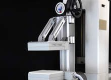
Hruska’s team set out to use the updated scintimammography technology, which they called molecular breast imaging, to see how well they could detect small cancers. They started using detectors on just one side of the breast, but soon introduced another set of detectors, so there were detectors above and below the breast. The equipment was supplied by industry collaborators GE Medical Systems from Haifa, Israel, and Gamma Medica-Ideas in Northridge, CA, US.
“Women sit next to the machine and the breast is placed between the two detectors in a similar position to a mammogram,” explains Hruska. “The top detector moves down, lightly compressing the breast, just enough to keep the breast from moving during the exam. As the imaging is picking up the tracer, any movement will likely smear the image. However, MBI does not require the high compression forces used for mammography; it is supposed to be a pain-free examination.”
At the moment, four views are taken – two of each breast – each lasting about ten minutes, so the full examination takes about 40 minutes. The team are working to reduce the time to under 20 minutes, but as Hruska explains there is always a trade-off. “Although a higher tracer dose would allow the imaging to be done more quickly, the team prefers to give patients a smaller radiation dose and take longer,” she says.
Patient studies
The first study, with 200 patients in 2001, used a single-head detector to see if it was possible to detect breast cancer. The patients all had suspicious lesions identified by either mammography or ultrasound, as well as a scheduled biopsy so that the team could compare their findings against a definitive result.
‘Although we had really good sensitivity with those initial studies detecting most of the cancers, we were still missing cancers that were very small, for example under 5mm, and cancers in women who had very large breasts,’ says Hruska. ‘Also, some of the images were count-poor – they didn’t have enough gamma rays collected in them to make an accurate assessment.’
The first study was followed by another using the dual-head detector system. Having two detectors reduced the maximum distance that a lesion or tumour could be situated away from either detector. This improved the sensitivity. At the same time the team made some technical improvements to the collimator, which filters and aligns the gamma rays to improve the count. Following this second study it was clear that molecular breast imaging could reliably detect smaller cancers.
The team then moved on to a large MBI screening study, in which 1,000 women were enrolled, and the results were impressive. “All these women were asymptomatic and coming in for their annual screening mammogram,” says Hruska. “They had a mammogram and then they also had the MBI test. We ended up detecting ten cancers, whereas only three were detected on mammography. We also had a lower number of false positive results than mammography as well, so fewer unnecessary biopsies.
“We’re still going through the data because we have to follow up these women for an additional 15 months to see if any more cancers will occur among the 1,000 women in that time frame.”
Imaging challenges
As promising as MBI is, the team still faces many challenges. Although the tracer used was not designed specifically for cancer detection, the team has seen it taken up by a variety of cancers including pre-cancerous lesions, invasive and ductal cancers. They have detected ductal carcinoma in situ, an early-stage cancer, and lobular cancer, which is sometimes harder to detect on mammography. However, the team will be rolling out studies with markers that are specifically targeted at cancer detection.
The main challenge at the moment is to work on methods to reduce the radiation dose, as this will be essential for MBI to move from being a diagnostic tool to become an effective general screening tool. Hruska believes it will eventually be possible to get the radiation dose down to the same level or lower than conventional mammography. There will also be improvements to the comfort and appearance of the machine.
Trials are also planned or underway, including a low dose trial, one looking at MBI as a pre-operative evaluation tool just prior to surgery to see if it can pick up additional cancers, and also a trial to evaluate the use of MBI for monitoring the response of tumours to chemotherapy.
As well as the quality and accuracy of the detection on MBI, the technology also has the advantage of being relatively inexpensive. Potentially it lends itself to use as a widespread population screening tool as well as a further diagnostic tool, given that its eventual cost is expected to be comparable with a digital mammogram and considerably less expensive than MRI.
Hruska is optimistic about the future for MBI technology, and the advances in medical imaging it is likely to herald. “The concepts of functional imaging have been there, but we didn’t have the technology to do it,” she says. “Now we are at a crossroads where much of the technology required is coming together.
“I feel that in medical imaging, even beyond the breast, the concept of looking at molecular tracers or cellular activity rather than just looking at the anatomy of the body is going to be the future of imaging.”
Crucially, these advances in medical imaging have practical social benefits. “It’s very rewarding and very exciting,” says Hruska. “Just take our screening study: we had a number of patients where we detected their cancer.
“If they hadn’t enrolled in the study they would have been sent home with breast cancer thinking they had a mammogram that was negative and that they were fine. These are things that really made a difference in these people’s lives.”
