According to the World Health Organisation, every year around 15 million people worldwide suffer some form of stroke. Of those that survive, many find themselves strangers to a new reality of disability and dependency, adapting to personal circumstances that have fundamentally and permanently changed.
Building empathy for a condition so pervasive in its impact shouldn’t be hard for a country like the UK, known as it is for a comprehensive and universal health system. But stroke is, at least historically, something of an anomaly for the British people. Their attitudes towards what should be a shared fear are notable for misinformation, negativity and a remarkable sense of common indifference.
Correcting neglect of this kind would be difficult without at least some consideration of the underlying cause. And though intuition may be the only tool available in making judgements, some sound explanations do exist. By far the most reasonable theory around is that the attitudes we show reflect an underlying resignation to stroke being a fact of life and a condition of old-age.
In 2008, the UK Department of Health commissioned the development of a National Stroke Strategy (NSS) to reverse the kind of unconstructive fatalism that typified this entire way of thinking.
Speed is key
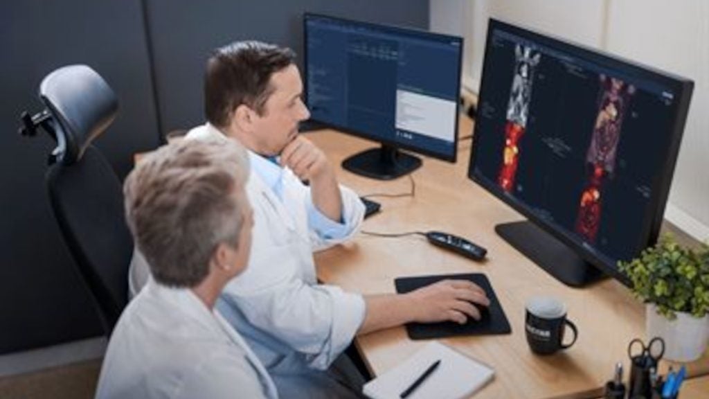
One of the major recommendations made by the strategy was the importance of fast, clear and case-specific imaging in the diagnosis of stroke. It’s now well established that diagnostic scanning can provide precise visuals of individual cerebral malfunction, information that is required for the safe prescription of life-saving drugs. Having this quick, early means of establishing the problem is crucial if the interventional stage is to shake off its laggard and ineffective past.
Of course, things haven’t always been this clear. At the time when Professor Anthony Rudd, consultant physician and chair of the College of Physicians Intercollegiate Stroke Working Party, was originally asked to draft the national stroke guidelines, there was almost no evidence to suggest that brain scanning might be useful in diagnosing strokes.
How well do you really know your competitors?
Access the most comprehensive Company Profiles on the market, powered by GlobalData. Save hours of research. Gain competitive edge.

Thank you!
Your download email will arrive shortly
Not ready to buy yet? Download a free sample
We are confident about the unique quality of our Company Profiles. However, we want you to make the most beneficial decision for your business, so we offer a free sample that you can download by submitting the below form
By GlobalData"When I first sat round a table with the intercollegiate working party drafting the first guidelines back in 2000, it took quite a lot to persuade the group to come to a consensus that brain imaging was needed at all in stroke care," he recalls.
Since then, things have changed a lot and imaging is now well known for its diagnostic credentials. At the forefront of this change has been the growing use of computed tomography (CT) scanners as the primary evaluative tool in the acute setting. Its popularity owes much to the clear way it differentiates between haemorrhagic and ischaemic stroke, as well its accessibility.
CT or MRI?
The importance of CT scans in stroke diagnosis has remained consistent over the past two decades despite the introduction of MRI onto the medical scene, but in recent years the principles that guided that consensus have changed.
Since the diagnostic potential of MRI was first mooted, research output has been steadily rising. The academic consensus coming out of those studies is clear: MRI outperforms CT in diagnosing patients suffering an acute stroke.
Dr Elizabeth Brown is a consultant radiologist at Gloucestershire Royal Hospital who was present at a key meeting of the Royal Society of Medicine last year. She argues
that the growing interest in MRI’s diagnostic capacity is well founded.
"It’s imperative that the diagnosis of stroke is confirmed very rapidly," she says. "We need to know what type of stroke it is and whether or not there’s an alternative verdict. Some strokes are difficult to see for up to several hours, even with the best scanner. With MRI, we’ve realised that you can actually detect what we call thrombotic stroke within minutes."
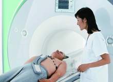
The strength of the magnetic fields produced by MRI scanners is capable of creating more conspicuous 3D images in far greater detail than other devices. This gives radiologists a more accurate picture of the exact problem, particularly when the issue involves inestimably small legions. Adding this level of subtlety to the diagnostic stage means more safety and more accuracy to the management process. What MRI therefore offers is not just a better capacity for diagnosis, but a safer way of orchestrating the treatment procedure.
To the general public and to medical professionals, results like these seem very exciting, but Brown cautions that we need to retain a sense of perspective.
"There are still many good reasons why CT remains the primary workhorse," she says. "If the patient is ill and unstable, the MRI environment is really not a good place to be. And aside from being unnerving, there are still a lot of contraindications to people going into the MRI scanner. A lot of the monitoring equipment used in the treatment of sick patients is incompatible with the large magnet that MRI scanners require."
Aside from proclaiming the benefits of MRI, many medical practitioners think of it as a replacement for CT. Brown is not so sure. For her, the purpose of MRI is not to displace existing technologies, but to broaden the scope of what’s available.
"Politicians always say that this is going to replace the previous scanner, but really this just gives us another weapon in the armamentarium, a better range of tools to do the best job for any individual patient."
To some extent, Rudd agrees on this point: "We are in a situation where we do need to be responsible about the way we use the resources we’ve got. As a stroke physician, I would argue that MRI is not needed for everybody. One could make a clinical justification for doing MRI probably for around half the patients with strokes."
MRI’s impact on neuroimaging
Whether we should view MRI as a replacement or a complement is an area that requires further discussion. There is however, no real question about its overall benefits. Since raising the profile of MRI scanning in stroke diagnosis, clinical outcomes have unambiguously improved. The last round of data from the national sentinel stroke audit team, a report co-authored by Rudd, illustrates a straight decline in the mortality rate from 24% to 17% between 2004 and 2010. Whether or not that improvement is down to MRI is difficult to establish, but it does seem likely to have contributed something.
"Picking out the data to see precisely what MRI has contributed would be virtually impossible," says Brown. "I think working out what’s causing the improvement is a combination thing."
Rudd shares precisely that sentiment: "I think it will be playing a part but I can’t say the percentage fall in mortality is entirely attributable to it," he says. "It’s really the complete package of managing strokes, taking the disease seriously, accepting that it’s important and knowing what causes it."
Having established the importance of MRI in diagnosing strokes, perhaps the most pressing concern for medical professionals and government actors is finding ways of fulfilling its potential. But, perhaps predictably, a consensus here is almost impossible to find.
For Brown, the chief practical problem with using MRI technology in the acute setting is operational pressure. Unless the strain placed upon these scanners by already well-utilised radiology departments is tackled, stroke patients will not have the straightforward access to MRI that they need.
"All these plans for early imaging and supporting rapid access to the scanner don’t, in my experience, seem to have come with a lot of money," she argues. "It just hasn’t really been financed. What I think we need is to improve the number of scanners available"
According to Brown, without this extra investment, the UK Government has some serious thinking to do. It’s generally accepted that in the past two decades central government has been relentlessly managerialist in its policy towards health, with external targets and waiting lists prioritised over good common sense. For Brown, today’s government must face up to this challenge. Does it want to retain its fidelity to waiting lists? Or does it want to offer an acute service on demand?
"The government wants it both ways," she says, "but it’s just not possible with what we have."
Rethinking resources
Rudd sees things differently. For him the point is to make much better use of the equipment already available.
"I think, with a judicious use of the resources we already have, we can get everything balanced far better," he says. "If you look at the way we use our scanners, most are lying idle over weekends and at night, so I would argue that there isn’t a justification for buying more scanners. Individual sites may have particular demands that do require it but in most cases we don’t need it."
One aspect on which the two certainly do agree is that any attempt to step up the use of MRI will require a thoroughly funded, intensive training programme for medical staff. Having the appropriate reading skills to interpret MRI scanners is not something all radiologists have in equal measure. For the time being, extra investment in training and equipment will be hard to find. As the country enters the start of a long age of austerity, making better use of the resources already available may be the only option available.
With or without financial support, deep changes have already been made both in the contribution of MRI imaging to stroke care and in broader attitudes towards the disease. Since the first strategy was published nearly a decade ago, improvements to stroke care have been dramatic. Many of the problems that do still remain are not impossible medical complications but practical dilemmas with practical solutions. Attention should now be drawn towards solving those issues and using the technology we know can make a difference.

This article was first published in our sister publication Medical Imaging Technology.


