
Alzheimer’s disease is devastating. Thirty million people suffer from dementia worldwide, and by 2050 this figure is predicted to rise to over 100 million. In Caucasian populations, Alzheimer’s disease accounts for over 50% of dementia cases. There is no known treatment, the causes of the disease are not known for certain, and the ability to run effective trials for possible treatments is complicated by that fact that diagnosing early-stage Alzheimer’s is difficult.
One popular theory is that the development of Alzheimer’s disease is linked to the aggregation of amyloid beta (Aß) deposits and the presence of neurofibrillary tangles formed by hyperphosphorylated tau proteins. Assessing the accumulation of these deposits is difficult, requiring histological assessment of tissue samples obtained by brain biopsy during the patient’s lifetime or, as is usual, on post mortem.
Go deeper with GlobalData

Discover B2B Marketing That Performs
Combine business intelligence and editorial excellence to reach engaged professionals across 36 leading media platforms.
A team of research scientists, including Juha Rinne, principal investigator for PET studies, Turku PET Center; Hilkka Soininen, Tuula Pirttilä, Irina Alafuzoff, Mikko Hiltonen and Anne Koivisto, University of Kuopio, Finland, is developing a less invasive method of detecting Aß in the brain using positron emission tomography (PET) scanning. Amyloid precursor protein (APP), a trans-membrane glycoprotein, is believed to play an essential role in maintaining the function of brain neurons. But when APP is broken down through sequential cleavage, it can create various isoforms of beta amyloid, notably Aß40 and Aß42.
It is Aß40, the less common of the two, that is implicated in Alzheimer”s through its formation of clumps of Aß outside the neurons, known as plaques, interfering with their proper functioning.
“The link between Aß and Alzheimer’s has been supported by plenty of studies,” says Dr Ville Leinonen, lead researcher at the University of Kuopio.
“Most of the research suggests that in the first phase there is some accumulation of Aß, and then over time the amyloid plaques get condensed. Other plaques are formed and, when they are present in the right concentrations, then you get the clinical disease.”
The Aß accumulation does not occur all at once, but progresses systemically through the brain. In the first phase, Aß deposits form at the margins of the brain in the neocortical regions and progressively works through the central regions towards the base of the brain. So it is present in the allocortex (second phase), diencephalic nuclei and striatum (third phase), distinct brainstem nuclei (fourth phase), and the cerebellum and additional brainstem nuclei (fifth phase).
“One of the most interesting aspects of Aß is that researchers are pointing to it as a precursor of Alzheimer’s disease,” says Leinonen. “If you find Aß early enough, you can find patients who may need prophylactic therapy before they develop any symptoms.”
Lower invasive risk
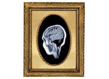
To help develop potentially promising treatments for Alzheimer”s disease and run clinical trials, it is important to develop a reliable means of detecting Aß that is less invasive than brain biopsy. One possible route to achieving this is to view Aß aggregation in the brain using PET with N-methyl-[11C]2-(4′-methylaminophenyl)-6-hydroxybenzothiazole, a compound derived from thioflavin T, as an imaging agent. This compound is better known as Pittsburgh Compound B (PiB) after the University of Pittsburgh, where research scientists William E Klunk and Chester A Mathis, originally identified it.
“We realised that there might be a chance to study the reliability of the PET imaging,” says Leinonen. “When we started the project there was no human study comparing the amyloid histology in brain samples with the results of PET imaging: we had the right facilities and the right patients to make that study.”
To test whether or not PET imaging could effectively detect the presence of Aß, the researchers needed to compare the results of the [11C]PiB PET imaging with the results of patients who had been subjected to a brain biopsy.
Tracing plaques
Patients with normal pressure hydrocephalus (NPH) exhibit the classic triad of symptoms: dementia, gait disturbance and urinary incontinence. Diagnostic tests include intracranial pressure (ICP) monitoring and a biopsy for a differential diagnosis. Complications from this procedure have been low, and the test is useful: in studies, 22–42% of patients with symptoms suggestive of NPH have shown Alzheimer’s disease pathological lesions in frontal cortical samples.
Between January 2004 and February 2007, 125 patients underwent ICP monitoring with frontal cortical biopsy for suspected NPH at the Department of Neurosurgery at Kuopio University Hospital. After an independent neurologist screened the medical records of the patients who were 75 years or younger, 50 patients were found to fulfil the study criteria. Ten of these patients agreed to participate in the [11C]PiB PET study.
These patients then underwent the procedure for monitoring intracranial pressure. A right frontal 12mm burr hole was made under local anaesthesia, and before the intraventricular monitoring catheter was inserted, brain biopsy specimens of 2-5mm in diameter and 3-7mm in length were obtained. Histological examination showed that six patients had Aß pathological lesions, and three also had hyperphosphorylated tau. Four patients had no Alzheimer’s disease-related pathological lesions in
the biopsy specimen.
Prior to their PET scans, patients were tested for cognitive impairment with a range of standard tests, including the Clinical Dementia Rating sum of box score, Mini-Mental State Examination, and Consortium to Establish a Registry for Alzheimer’s Disease Neuro-psychological Test Battery. In the PET investigations, the radiochemical purity of the [11C]PiB tracer that was injected was more than 98%.
Patients had a 90-minute dynamic PET scan with a GE Advance PET scanner in the 3D scanning mode. Analysis of the tracer uptake in different areas of the brain was carried out with whole brain voxel-based statistical analyses using statistical parametric mapping and automated region of interest (ROI) analysis of regions bilaterally on the frontal cortex, lateral temporal cortex, medial temporal lobe, inferior parietal lobe, occipital cortex, cerebellar cortex and subcortical white matter.
Compared with those patients that had no notable Aß aggregates in the brain biopsy specimen, the patients with Aß aggregates in the frontal cortex showed significantly higher uptake of the PiB tracer in the frontal, parietal and lateral temporal cortices (phase 1 of regional Aß deposition) and striatum (phase 3).
With the automated ROI analysis, compared with those patients that had no notable Aß aggregates in the brain biopsy specimen, the patients with Aß aggregates in the frontal cortex showed significantly higher uptake of the tracer in the lateral frontal and lateral temporal cortices (phase 1), anterior and posterior cingulate gyri (phases 2 and 3), and caudate nucleus (phase 3). Patients with the highest Aß load from the biopsy, had the highest uptake of tracer.
Encouraging findings
The findings were encouraging regarding the future use of [11C]PiB PET imaging for the non-invasive evaluation of Aß deposition in patients, although further studies are required to see how useful this imaging method will be as a tool for diagnosing Alzheimer’s disease. It also shows promise as a means for monitoring patients undergoing treatment related to the accumulation of Aß in the brain.
“It might be reasonable to study patients with mild early symptoms,” says Leinonen. “That would be a population that might be good candidates for scanning to see if they have positive PET scan and are suitable for trials of some kind of amyloid-blocking therapy.
“There is a small dose of radiation because of the radiation agent, but the risk is very small: the dose is less than a couple of CT scans. It is a one-day visit that includes an MRI scan and a PET scan.”
However, there are still challenges to deal with. For example, it may be that the time between the biopsy and PET scan skewed the results. False negatives might be possible due to the small biopsy sample. There was one case where the tissue analysis was positive for Aß, but the PET scan negative suggests that it might be possible that there are types of Aß deposits that are not detected by [11C]PiB PET imaging.
“If the PET scan is positive we are more confident that the Aß is present,” he says. “In most cases when the PET scan is negative we believe it is truly negative, but there are some situations where Aß might not be detected by this PET scan. So we can’t say the PET scan is 100% effective, but it is 90% at least. It’s the best non-invasive method. However, we still don’t know for sure that, say, if a patient aged 70 happens to have Aß in the brain, that this will inevitably lead to Alzheimer’s disease in the next ten years. We still have to find this out.”
