
Having recorded its first X-ray image less than a year after the discovery of X-rays, Shimadzu has been at the forefront of medical imaging, as Matthias Schmidbauer, medical devices consultant for Shimadzu Europa explains.
For more than 100 years, Shimadzu has been counted among the most innovative companies in medical technology. Merely ten months after the discovery of X-rays, Shimadzu recorded the first X-ray image in Japan. To this day, the company is one of the pioneers in diagnostic imaging.
Recommended Buyers Guides
In recent years, the medical systems division achieved the largest growth rates. Within the company Shimadzu focuses extensively on direct radiography systems and has introduced additional novel diagnostic and interventional radiology products to the European healthcare market.
An essential element of Shimadzu’s product range is the Safire flat-panel detector, which is based on the principle of direct conversion. This new technology has been further developed for radiography, fluoroscopy, angiography and cardiology systems.
Ground-breaking detector technology
The Safire direct-conversion flat-panel detector offers excellent image quality with an outstanding detailed resolution. Safire converts X-rays directly, without any intermediate steps, into electrical output signals that can be digitally processed.
Conventional image amplifiers or flat-panel detectors that work according to the principle of indirect conversion need several intermediary steps in order to generate an output signal. However, each step also generates an interfering signal (noise). The original signal component in the output signal decreases with each step and the signal-to-noise ratio deteriorates. This actual loss of information can only be compensated by a higher radiation dose – this, however, burdens the patient. The benefits of the Safire technology are therefore obvious: based on its higher quantum efficiency, it is possible to achieve a higher resolution at a defined radiation dose or a defined resolution with a lower radiation dose.
The Safire flat-panel detector, 600 of which have been installed to date, is available as a 9in (23cm x 23cm) and 17in (43cm x 43cm) version.
Reduced dose and optimised image quality
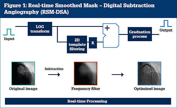
Using the Real-Time Smoothed Mask Digital Subtraction Angiography (RSM-DSA) technology patented by Shimadzu, filtered reference images are continually subtracted from the original images and from these data optimised images are then calculated and represented in real time (see Figure 1).
Thanks to this procedure, DSA-typical motion artifacts (breathing, heartbeat or patient-induced movement) as well as the noise component in fluoroscopy images are significantly reduced. Multiple-bolus dosing, essential in conventional systems, is superfluous using RSM-DSA.
High-resolution fluoroscopy images can be generated within shorter patient examination times – all this at lower radiation doses and lower contrast agent use.
The SUREengine real-time image processing software package
The SUREengine is a real-time image processing software package for the Safire flat-panel detector, specifically designed for image improvement and dose reduction.
For digital data processing the frequency spectrum is first decompounded into wave bands. The noise components are then subtracted from the individual frequency bands, and the image contrast is selectively increased. In this way, for instance, guide wires and catheters become much more clearly distinguishable.
In order to broadly reduce blooming and to display evenly illuminated image backgrounds, the entire dynamic range is compressed. Moreover, Moiré patterns resulting from scattered radiation grids are filtered out of the original images without any loss in image quality.
State-of-the-art diagnostic imaging technology
With the introduction of the Safire technology, Shimadzu has opened the gateway to new applications such as digital tomosynthesis, slot radiography, dual-energy subtraction and CT-like imaging.
Digital tomosynthesis
Digital tomosynthesis is the next-generation development of imaging tomography. During a sweep, the flat-panel detector records up to 90 images from different camera angles. Two calculation algorithms are subsequently used for data processing:
- The so-called shift integration, whereby stacked individual images are compiled, merged and layered and are then reconstructed
- The well-known CT back projection method
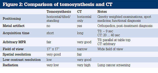
Unlike other sectional imaging techniques, tomosynthesis can also be applied to patients in an upright position. This is particularly helpful during exercise testing on joints such as hips and knees, or gravity-weighted examinations.
The advantages of direct radiographic tomosynthesis in comparison with computer tomography are considerable. Based on the lower patient radiation dose and the shorter physical examination time required, this procedure is suitable in particular for functional and orthopaedic diagnostics and for lung cancer screening (see Figure 2).
Slot radiography
During slot radiography a sequence of 5cm wide, adjoining bands (slots) is acquired using almost closed collimator geometry and pulsed radiation, while X-ray tubes and flat-panel detector move linearly in one direction.
After examination, the image bands are digitally reconstructed into a so-called long image (leg / spine). Therefore, this procedure is used in scoliosis and orthopaedics examinations, in order to completely map the spine or the lower extremities.
The component of the elicited image noise due to scattered radiation is significantly reduced in slot radiography. The use of a grid is no longer necessary. The image contrast is improved and the patient radiation dose can be decreased.
Dual-energy subtraction
Dual-energy imaging enables fast image acquisition using alternating high and low value kV-pulses (e.g. 140kV and 60kV).
During post processing, the resulting images are subtracted from each other and a soft tissue image as well as a bone image can be calculated after previous standardisation with respect to bone or lung values.
The main advantage of this procedure is the clear representation of calcifications, which is extremely helpful in the detection of lung nodules.
CT-like imaging
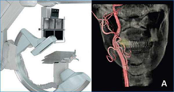
CT-like imaging closes the gap between angiography and computer tomography systems in interventional radiology.
The digital data sets acquired during C-arm rotation can be reconstructed – similarly to computer tomography – to represent delicate three-dimensional vascular structures (Figure 3).
In comparison with a 128-line CT, CT-like imaging examinations using a flat-panel based C-arm can lead to resolution improvements of up to 200%, while the patient dose can be simultaneously reduced by 90%.
This technology is frequently applied in interventional radiology and for tumour localisation in cancer therapy.